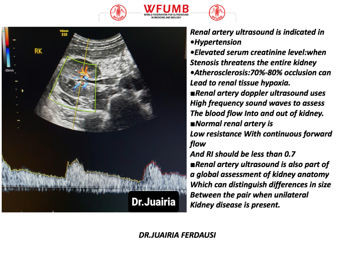


If your test results show abnormalities, including indicating the presence of PAD, your vascular doctor will be able to recommend appropriate follow-up care and treatment. Ultrasound does not use ionizing radiation and has no known harmful effects. It is commonly used to search for blood clots, especially in the veins of the leg a condition often referred to as deep vein thrombosis. It can show blocked or reduced flow of blood through narrow areas in the major arteries of the neck. It helps doctors assess the blood flow through major arteries and veins, such as those of the arms, legs, and neck. Using ultrasound technology and blood pressure cuffs, your doctor will be able to detect any narrowed or blocked blood vessels, as well as any arteries that have abnormal blood flow. An arterial line was unable to be placed in 11 out of 66 patients (16.7) despite a Doppler ultrasound-trained physician being available. Venous ultrasound uses sound waves to produce images of the veins in the body. A Doppler ultrasound test uses reflected sound waves to see how blood flows through a blood vessel. Lower extremity arterial Doppler testing is noninvasive and painless. Your doctor at South Palm Cardiovascular Associates may use this test to detect and diagnose conditions including arteriosclerosis (plaque buildup in the arteries that causes peripheral arterial disease), deep vein thrombosis, venous insufficiency, or to evaluate injuries to the arteries. Knowledge and understanding of the embryologic basis of the renal vasculature are necessary for the radiologist. If your cardiovascular specialist suspects blood flow in your legs may be abnormal, he may assess your condition with arterial Doppler ultrasound. The physiologic role of the kidneys is dependent on the normal structure and functioning of the renal vasculature. at any gestational age, the diameter of the single umbilical artery is larger than the arterial diameter would be if there were two arteries 14.
#Arterial doppler ultrasound free
It can be carried out by a simple ultrasound scan. The decision to use a free loop of the cord was made early in the history of Doppler ultrasound and has been applied with great clinical success. Sound waves are emitted from and reflected back to the probe.
#Arterial doppler ultrasound skin
The Doppler probe is coupled to the skin with gel and angled to the direction of blood flow. Ultrasound Doppler estimates of femoral artery blood flow during dynamic knee extensor exercise in humans. Blood flow in the lower extremities (the legs) is often affected by peripheral arterial disease. A Doppler ultrasound measurement is used to check the blood flow in the uterine arteries. Procedures Continuous-wave Doppler: It applies the Doppler effect to moving blood red blood cells to assess flow velocity within a vessel. Arterial vascular ultrasound has, however, been hitherto relatively neglected in education.


 0 kommentar(er)
